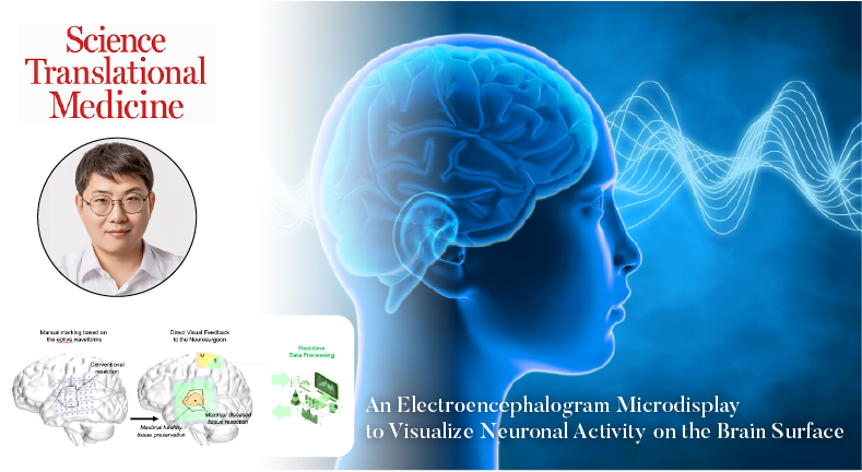
A groundbreaking technology has been developed for real-time brain activity monitoring, poised to revolutionize the success rates of brain surgery by enhancing precision and differentiation between normal and lesion tissues. According to the research team, this innovative technology allows for the real-time visualization of recorded cortical activity on the brain surface, offering immense potential for electrophysiological studies and neurosurgical procedures.
Led by Professor Youngbin Tchoe in the Department of Biomedical Engineering at UNIST, a research team has unveiled a revolutionary technology that enables the visualization of neuron activity on the brain surface in real-time. This innovation allows for the accurate removal of lesion tissues while safeguarding critical brain structures through the continuous monitoring of pathological activities during surgical procedures.
The intracranial electroencephalogram (iEEG)–microdisplay, pioneered by the research team, has the ability to measure, interpret, and visually present brain cortex activity in real time. This ingenious tool plays a vital role in identifying boundaries with utmost clarity by electrophysiologically differentiating and illustrating essential brain tissues during surgical interventions.

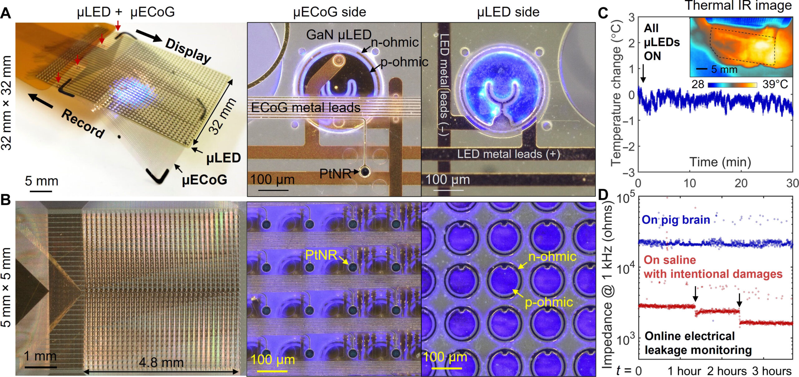
Figure 1. Fabrication process and safety evaluation of the iEEG-microdisplay.
Precisely delineating functional boundaries is crucial when excising specific brain areas affected by lesions during surgery. The iEEG–microdisplay, created by thousands of micro-ECoG channels with thousands of flexible microLED pixels, facilitates real-time visualization of brain activity, enabling neurosurgeons to perform successful operations with enhanced precision.
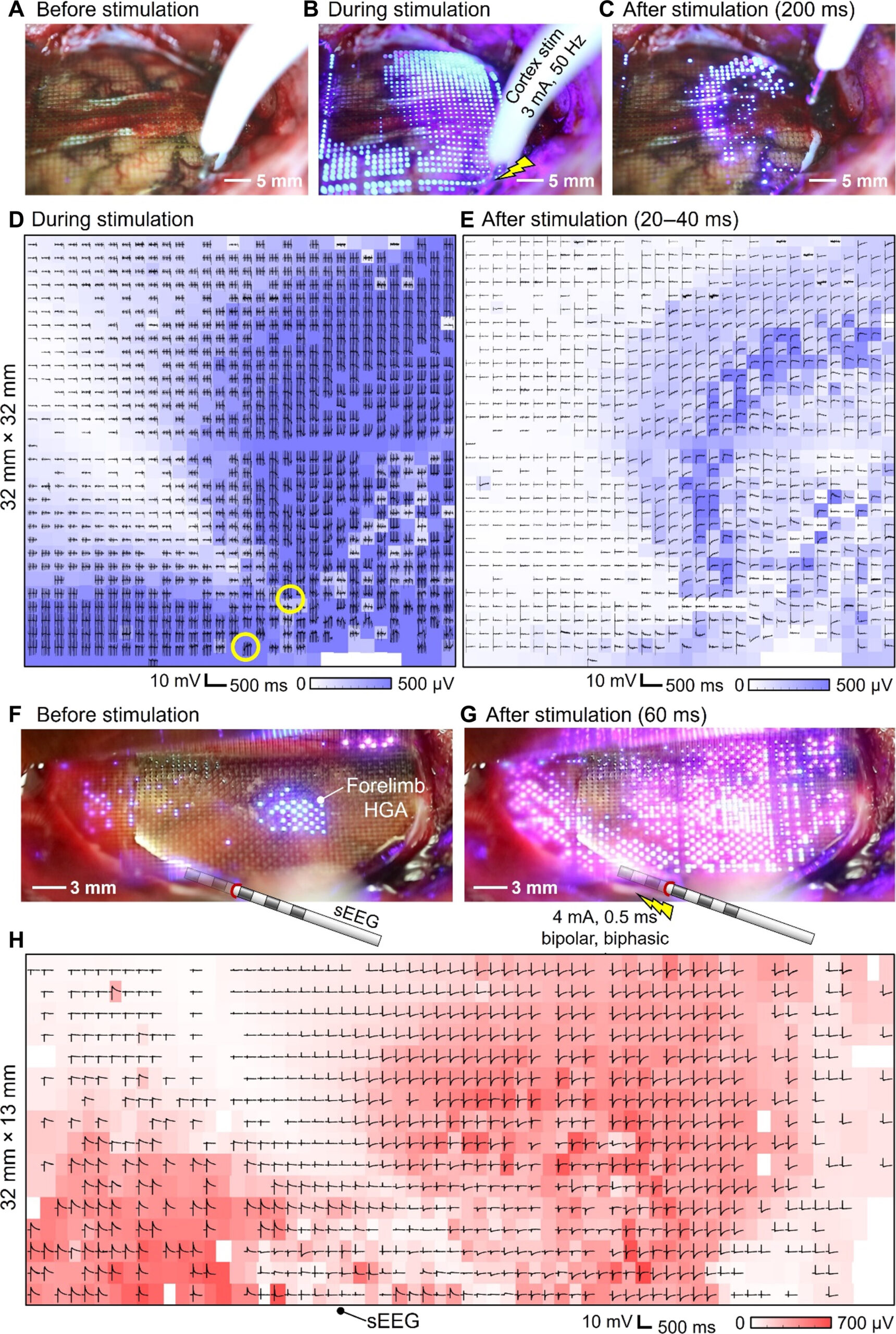
In validating the efficacy of this cutting-edge technology, the research team conducted experiments on pigs and rats. The team’s pioneering work is anticipated to make significant contributions to advancements in neurosurgery by providing real-time insights into the structure and function of the brain through brainwave display.

Figure 2. Display of electrical potentials in response to DES on the cortical surface of pig brains.
Professor Tchoe expressed his excitement over the technology’s potential, stating, “This development allows us to visualize the electrophysiological activity of the brain surface in real time during surgery.” Emphasizing the significant impact of this innovation, he highlighted its ability to pinpoint brain tissues requiring excision and those crucial for preservation, ultimately leading to improved outcomes for patients undergoing brain surgery.
The research findings have been published in the prestigious international journal Science Translational Medicine on April 24, 2024. This collaborative effort involved researchers from UC San Diego, Oregon Health and Science University, and the Massachusetts General Hospital (MGH), with support from the National Institutes of Health (NIH).
Journal Reference
Youngbin Tchoe, Tianhai Wu, Hoi Sang U, et al., “An electroencephalogram microdisplay to visualize neuronal activity on the brain surface,” Sci. Transl. Med., (2024).